Magnetic Resonance Imaging
 Magnetic Resonance Imaging (MRI) is a non-invasive technique that uses the magnetic properties of atomic nuclei (typically hydrogen) and nuclear magnetic resonance (NMR) signals to image the internal structure and workings of the body in vivo.
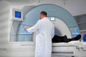
In a typical MRI scan, the subject lies inside a large cylindrical magnet that generates a static magnetic field of strength 1.5–7 Tesla, along the length of the scanner. The static magnetic fields align the atomic nuclei within the tissue of the subject. A rotating magnetic field then perturbs the static magnetic field, exciting the atomic nuclei to emit oscillatory radiofrequency signals. Those signals are converted via a receiver coil into electrical signals for encoding of the final MR image. Different body tissues have different magnetic resonance properties, and so produce distinct signals which enable imaging of the soft tissues of the body.
MRI is ideally suited for use in clinical trials to support
- disease understanding
- patient stratification
- diagnosis
- prediction of disease progression
- assessment of drug efficacy
HMR has facilitated MRI in clinical trials since 1993, and is proud to collaborate with the world-leading imaging and analytics service provider Invicro ( https://www.invicro.com) using their London-based infrastructure for clinical imaging services. MRI is used either as a standalone service or in combination with another imaging modality such as Positron Emission Tomography (PET).
Invicro London has 3 clinical MRI-capable systems: 2 dedicated Siemens MRI scanners, and a state-of-the-art combined GE SIGNA PET-MRI scanner. All the systems use a 3 Tesla magnet. Those MRI systems offer advanced MRI imaging, using a broad library of MRI techniques & sequences to study the structure and function of the body in both health and disease. Invicro London has outstanding expertise in brain applications of MRI in conditions such as Alzheimer’s, multiple sclerosis, depression, obesity, and pain, and is rapidly expanding into non-brain MRI applications such as osteoarthritis & rheumatoid arthritis, liver disease (including NASH), Crohn’s disease, and musculoskeletal conditions.
In a typical MRI or PET-MRI trial, HMR recruits the subjects, does the clinical work, and runs the trial. Invicro develops the MRI scanning protocol, does the MRI or PET-MRI scans, then analyses and interprets the results. We’ve used MRI or PET-MRI in more than 30 trials in healthy subjects or patients, to advance disease understanding or to assess novel IMPs.